|
|
Complex Regional Pain Syndrome
Reflex Sympathetic Dystrophy (Sudek Atrophy)
General Considerations
- Has also been called Reflex Sympathetic Dystrophy and Sudeck’s Atrophy
- Complex Regional Pain Syndrome (CRPS) overwhelmingly affects white females
- Cause is unknown but may involve an immune mediated mechanism
- Most cases occur secondary to fractures, sprains, and minor soft tissue injury
- Other causes include
- Head injury
- Stroke
- Myocardial infarction
- Cast/splint immobilization
- Upper extremities involved more frequently than lower extremities in adults
- CRPS Type 2 involves nerve injury and is called causalgia
Staging of CRPS |
Stage 1 |
- 0-3 months
- Puffy swelling, redness, warmth, hyperhydrosis, decreased ROM, radiographs usually normal, positive bone scan
- Aggressive treatment at stage 1 ® best outcome
|
Stage 2 |
- 3-6 months
- Worse pain, edema hardens, cyanosis, dry skin, worse stiffness, atrophy of skin, osteopenia on radiographs
|
Stage 3 |
- > 6-12 months
- Skin pale, cool, dry, tight and glossy; joint contractures
- Severe osteopenia with spindling of fingers; diminished pain; nails become rigid; hair becomes fragile
|
Clinical Findings
- International Association for the Study of Pain (IASP) criteria for diagnosis
Complex Regional Pain Syndrome Diagnostic Criteria |
The presence of an initiating noxious event or a cause of immobilization |
Continuing pain, allodynia (perception of pain from a non-painful stimulus), or hyperalgesia disproportionate to the inciting event |
Evidence at some time of edema (most common sign), changes in skin blood flow, or abnormal motor activity in the area of pain |
The diagnosis is excluded by the existence of any condition that would otherwise account for the degree of pain and dysfunction. |
Imaging Findings
- The diagnosis is made clinically
- Conventional radiographs may be normal (30%) or may show severe osteopenia about 2-3 weeks after the onset of the disease
- Classically has a sock-like distribution affecting distal tibia and foot but sparing remainder of the tibia
- Triple-phase bone scan
- Most sensitive (44%) earlier in disease (<20 weeks)
- Diffuse increased activity, with juxta-articular accentuation uptake on the delayed images (phase 3)
Differential Diagnosis
- Disuse osteopenia - missing the edema and pain associated with CRPS
Treatment
- Physical and occupational therapy
- Pain relief
- Sympathetic and somatic nerve blocks
- Antidepressants and anticonvuslants
- Transcutaneous electrical nerve stimulation (TENS)
- Lidocaine and ketamine have been used
Prognosis
- The earlier the disease is diagnosed, the better the prognosis
- Children seem to have a better prognosis than adults
- Late CRPS is refractory to treatment and could result in long-term disability

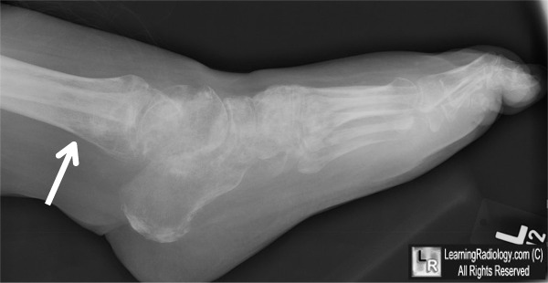
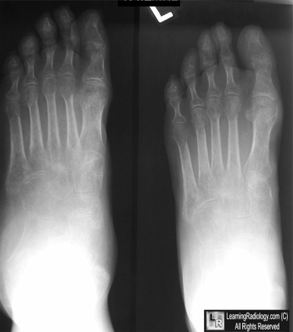
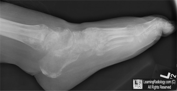
Complex Regional Pain Syndrome. Upper images show intense osteopenia involving the whole foot but more pronounced in a juxta-articular pattern. Lower lateral foot demonstrates the abrupt start of the osteopenia in the distal tibia (white arrow) and diffuse soft tissue swelling.
For these same photos without the arrows, click here and here
For more information, click on the link if you see this icon 
Complex Regional Pain Syndrome. eMedicine. S Parrillo.
|
|
|
